Clinical cases

Clinical cases, documented with care.

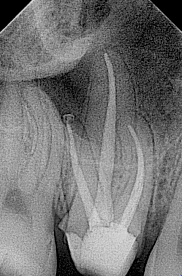
Side-by-side radiographs show the tooth before treatment, immediately after treatment when available, and at follow-up.

The radiographs below are de-identified and presented for patient education. Every tooth and healing response is different; these cases do not predict or guarantee an individual result. Select any image to view it at full size.

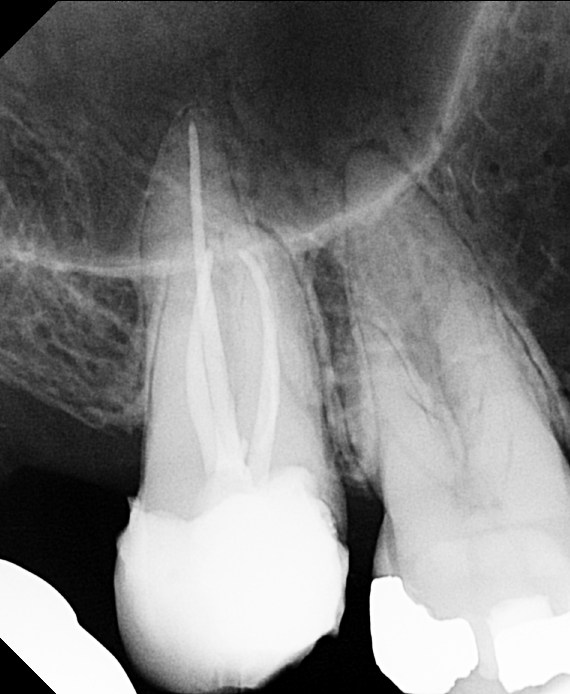
Case 01

Tooth #12
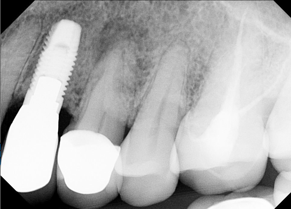
Six-month follow-up
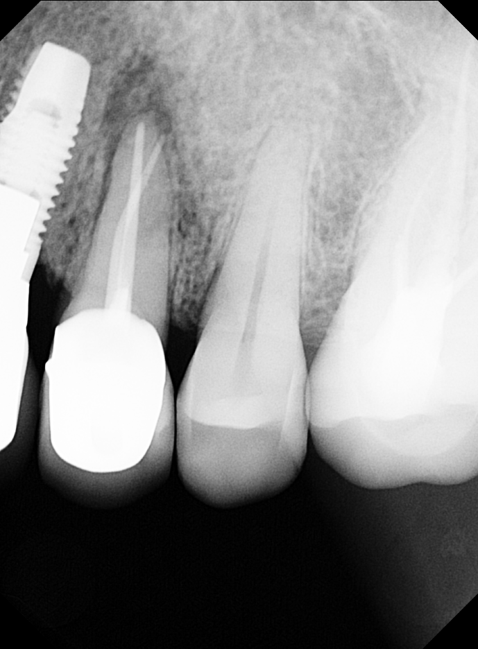
Pre-operative radiograph of tooth number 12
Pre-opBefore treatment
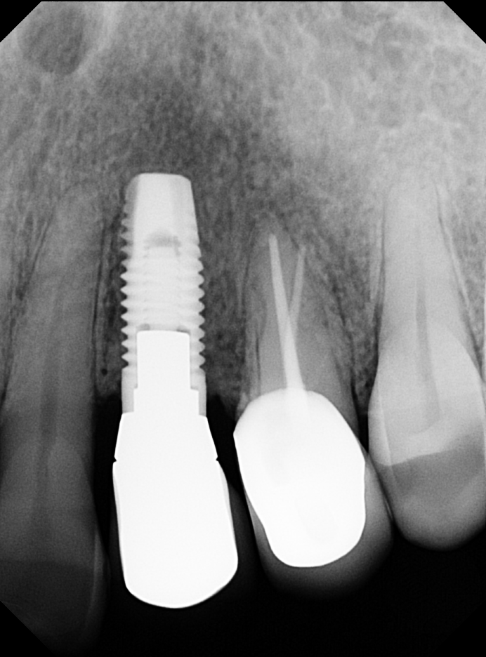
Immediate post-operative radiograph of tooth number 12
Post-opImmediately after treatment
Six-month follow-up radiograph of tooth number 12
Follow-upSix months

Six-month radiographic follow-up: The previously large root-end lesion shows bone fill and reformation of the periodontal ligament (PDL) space.

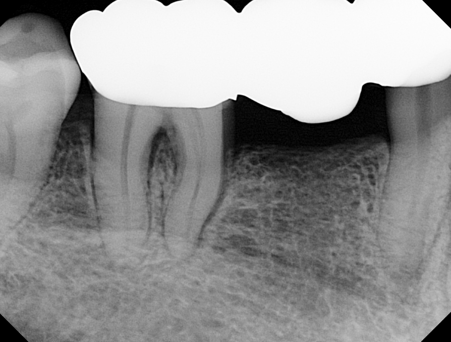
Case 02

Tooth #15
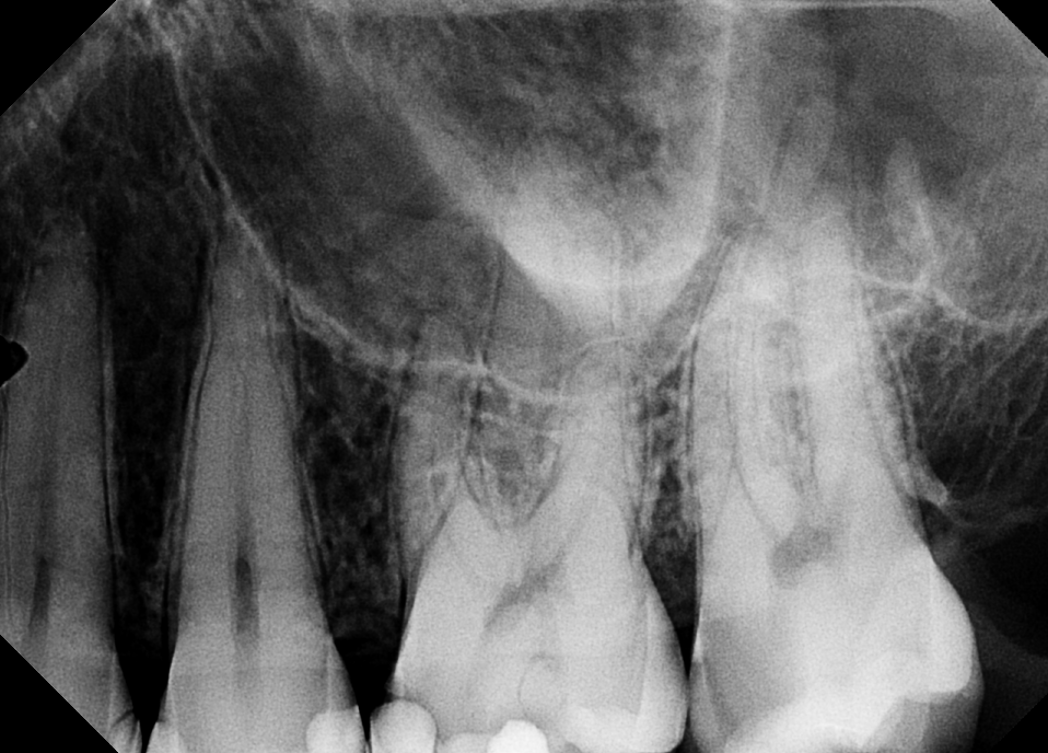
Pre-op · Post-op
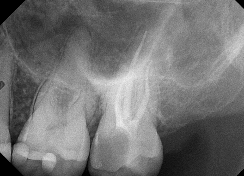
Pre-operative radiograph of tooth number 15
Pre-opBefore treatment
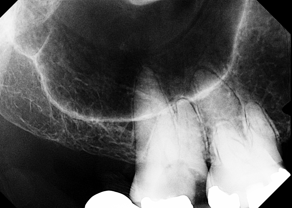
Post-operative radiograph of tooth number 15
Post-opAfter treatment

Case 03

Tooth #2

Pre-op · Post-op
Pre-operative radiograph of tooth number 2
Pre-opBefore treatment
Post-operative radiograph of tooth number 2
Post-opAfter treatment

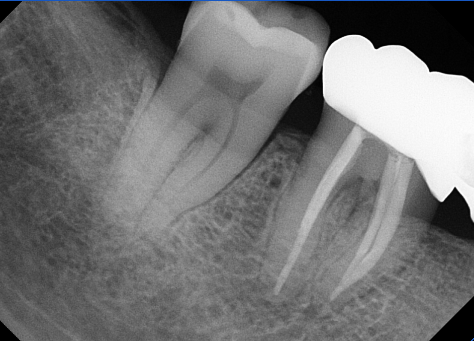
Case 04

Tooth #30

Pre-op · Post-op
Pre-operative radiograph of tooth number 30
Pre-opBefore treatment
Post-operative radiograph of tooth number 30
Post-opAfter treatment

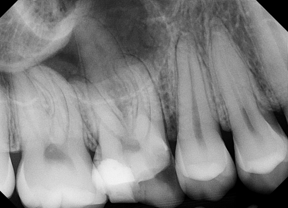
Case 05

Tooth #3

Pre-op · Post-op
Pre-operative radiograph of tooth number 3
Pre-opBefore treatment
Post-operative radiograph of tooth number 3
Post-opAfter treatment

Clinical images are de-identified and shared for patient education. Treatment recommendations and outcomes depend on each patient’s diagnosis, anatomy, restorability, and healing response.